Figure 1.
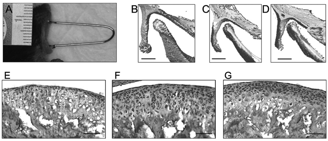
Forced mouth opening model. (A) Image of anesthetized mouse exposed to forced mouth opening. (B,C,D) The region of skull including the mandibular condyle was imaged by micro-CT (VivaCT40, Scanco Medical AG, Bassersdorf, Switzerland), with the mouth in the closed position (B) and then opened with spring forces of 0.25 N (C) and 0.5 N (D). Bar: 500 µm. (E,F,G) H&E staining of sagittal sections of the TMJ in 47-day-old female C57BL/6 mice exposed to 5 days of 0 (E), 0.25 N (F), or 0.5 N (G) forced mouth opening for 1 hr/day. Bar: 50 µm.

