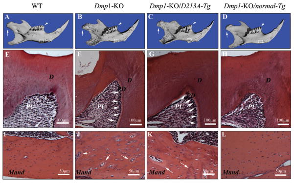
Figure 2.
Micro-CT images and H&E staining of the teeth and mandibles of the six-week-old mice. PD = predentin; D = dentin; PU = pulp; Mand = mandibular body. (A-D) Micro-CT images of the mandibles: WT, Dmp1-KO, Dmp1-KO/D213A-Tg, and Dmp1-KO/normal-Tg mice. The condyles were indicated by white arrows, the first molars analyzed by H&E staining were indicated by white arrowheads (E-H), and the mandibular areas analyzed by H&E staining were boxed (I-L). The three-dimensional micro-CT images showed that the mandibles of the Dmp1-KO (B) or Dmp1-KO/D213A-Tg mice (C) were shorter than those of the WT (A) or Dmp1-KO/normal-Tg mice (D). Compared with the WT and Dmp1-KO/normal-Tg mice (A, D), the surfaces of the mandibles in the Dmp1-KO or Dmp1-KO/D213A-Tg mice appeared more porous (B, C). (E-H) H&E staining of the mesio-cusp region of the first molars of six-week-old mice: WT, Dmp1-KO, Dmp1-KO/D213A-Tg, and Dmp1-KO/normal-Tg mice. Note that the predentin in the first molars of both Dmp1-KO and Dmp1-KO/D213A-Tg mice (F, G; white arrows) was wider than in the WT (E) or Dmp1-KO/normal-Tg (H) mice. (I-l) H&E staining for the mandibular bodies of six-week-old mice: WT, Dmp1-KO, Dmp1-KO/D213A-Tg, and Dmp1-KO/normal-Tg mice. Note that the mandibular bodies of both the Dmp1-KO and Dmp1-KO/D213A-Tg mice (J, K) contained more osteoid (areas indicated by white arrows) and enlarged osteocyte lacunae compared with both the WT and Dmp1-KO/normal-Tg mouse (I, L).

