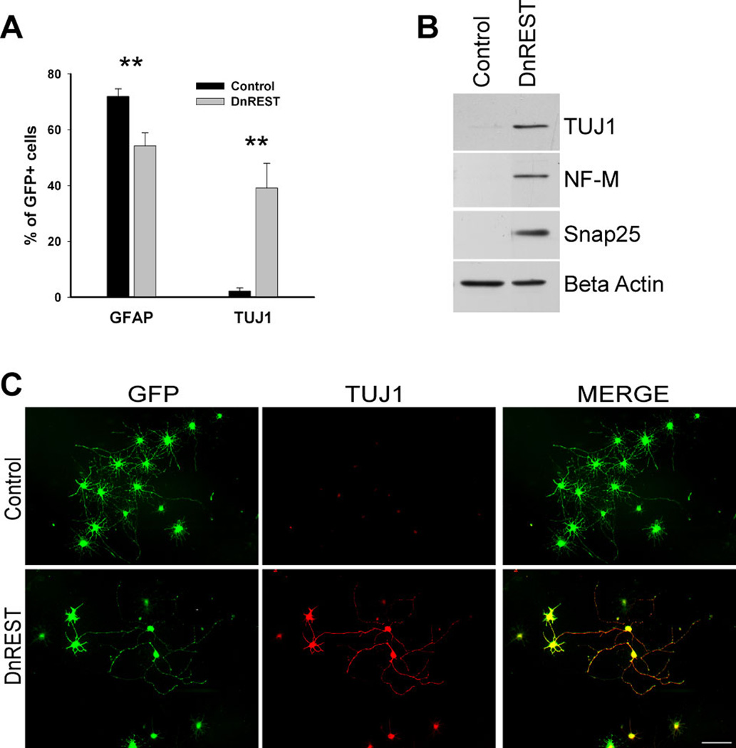
Figure 4. REST loss of function increases neuronal protein expression during astrocytic differentiation.
Purified OPCs were infected with retroviruses expressing either DnREST and/or GFP alone and grown for 5 days in 2A differentiation media. A. The percent of the infected cells that were positively stained with antibodies against GFAP and TUJ1. Error bars represent the standard deviation, n=3, ** P value < 0.001 B. Immunoblot analysis shows the expression of neuronal proteins in the DnREST infected cells after 5 days in 2A differentiation media. C. Immunofluorescence staining showing the expression of TUJ1 (red) in the infected cells (green) bar=50µm.

