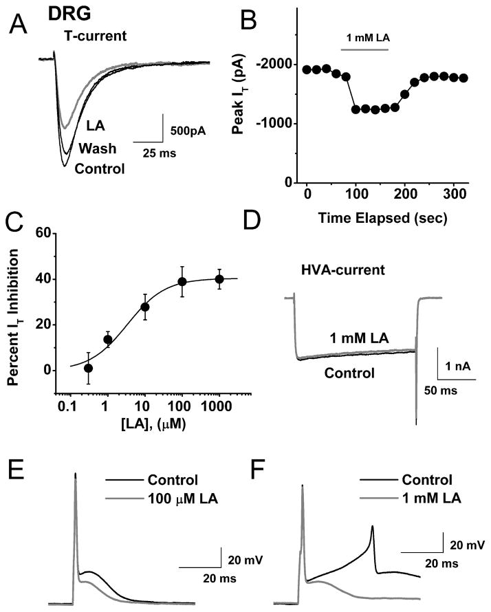
Figure 1. LA selectively inhibits T-type Ca2+ currents and underlying after-depolarizing potentials in acutely dissociated rat sensory neurons.
A. Traces of T-current in a representative DRG cell before and after (black traces), as well as during bath application of 1 mM lipoic acid (LA, gray trace), which reversibly inhibited about 38 % of peak inward current. Bars indicate calibration.
B. Temporal record from the same cell presented on panel A of this figure. Gray bar indicates duration of LA application.
C. Concentration-response relationship for LA inhibition of T-current in rat DRG cells (n = 6–20 per data point). Solid line is the best fit (equation # 1, see Methods) yielding IC50 of 3.3 ± 1.5 μM, slope coefficient 0.8 ± 0.2, and maximal inhibition of 40.4 ± 3.1 % of the peak of T-current.
D. Traces of HVA Ca2+ current from another DRG cell before (black trace) and during the bath application of 1 mM LA (gray trace). Bars indicate calibration.
E. AP waveforms in a representative DRG cell before (black trace) and during application of 100 μM LA (gray trace). Note that LA application had little effect on RMP and the initial AP spike, but attenuated the maximal amplitude of ADP by about 30% and the duration of ADP (measured at half-maximal height) by about 45%.
F. The same experimental protocol (described in panel E of this figure) was used in another DRG cell that fired a repetitive APs at the crest of ADP (black traces). When 1 mM LA was applied in the bath, the initial amplitude of ADP was decreased about 20% and the cell did not fire repetitively (gray trace) when the same single depolarizing stimulus was injected through the recording electrode.

