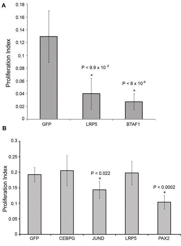
Figure 5.
Analysis of siRNA treatments in chick SC (SC) and retinal pigmented epithelia (RPE) proliferation. Percent proliferation was quantified for siRNA treatments in (a) chick SC for genes commonly down-regulated in treatments that inhibit SC proliferation (downstream of the AP-1 Pathway and CEBPG) and (b) chick RPE for genes that inhibited chick SC proliferation.

