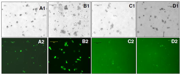
Figure 4. GFP expression in HSCs at day 10 of post transduction.
(A1, A2) bright field and florescence images of stem cells infected with EF1-GFP, (B1, B2) bright field and fluorescence image of stem cells infected with CMV-GFP; (C1, C2) bright field and florescence images of stem cells infected with SV40-GFP, (D1, D2) bright field and fluorescence images of stem cells infected with UBC-GFP.

