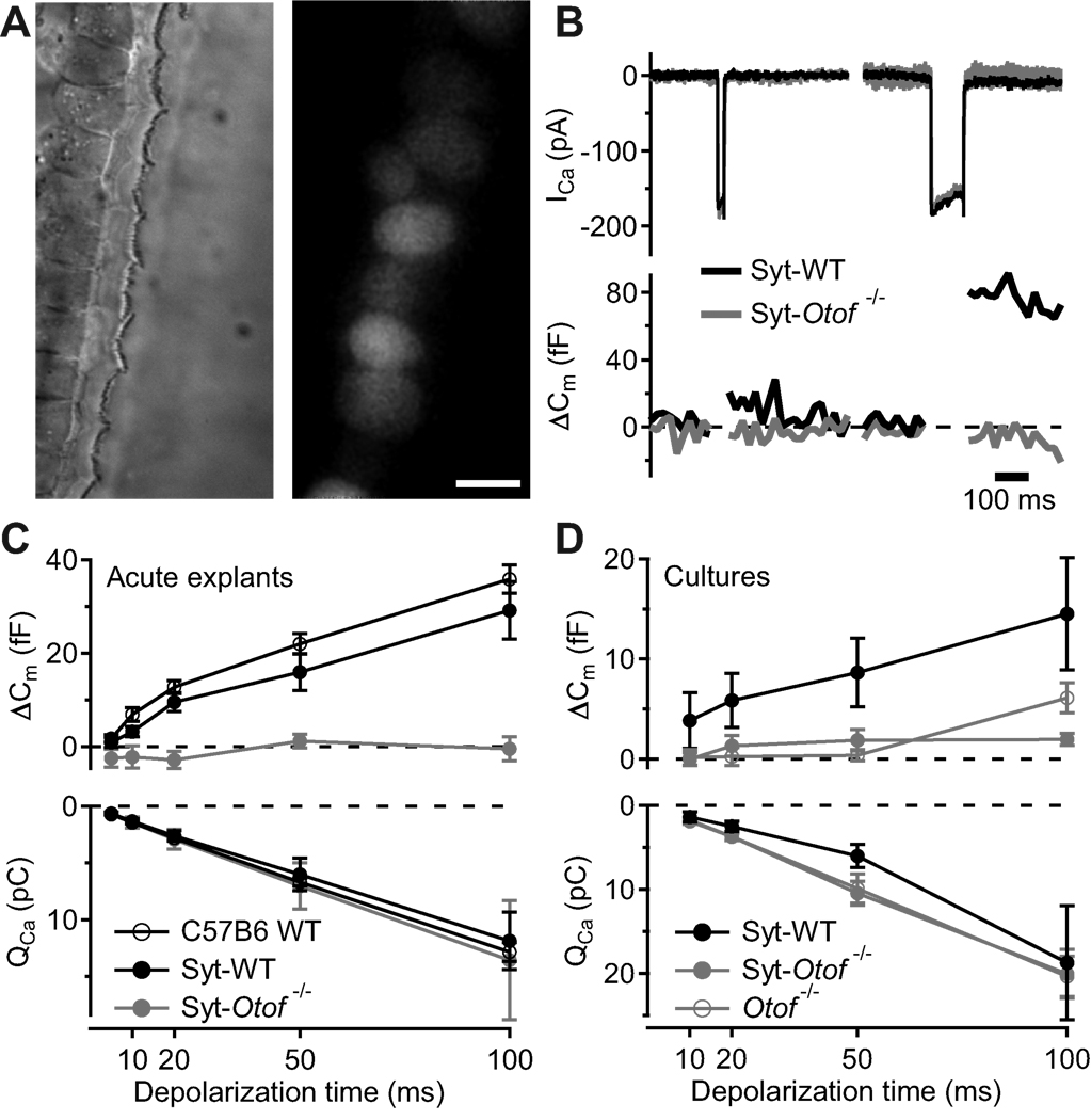
Figure 3. Syt1 overexpression by in vivo and in vitro AAV transduction do not restore exocytosis in IHCs of Otof−/− mice.
A, representative differential interference contrast (left) and fluorescence (right) images of p35 IHCs following transuterine transduction of the embryonic otocyst using AAV-1/2 Syt1-IRES-eGFP: preserved stereocilia and high transduction rate (chain of eGFP-positive IHC somata). Scale bar 5 µm.
B, synaptic function in Syt1 misexpressing IHCs in acutely isolated p35 organs of Corti. Representative ICa currents (top) and ΔCm in response to a 20 ms (left) and 100 ms (right) depolarization to −14 mV, recorded in perforated-patch configuration from IHCs of wild-type (black, CD1) and Otof−/− mice (grey, mixed background) that had been transfected in utero.
C, mean ΔCm (top) and QCa (bottom) responses of untransfected 4–week-old C57B6J IHCs (open circles, n = 12), AAV-1/2 Syt1-IRES-eGFP-transfected CD1 IHCs (filled black circles, n = 9) and AAV-1/2 Syt1-IRES-eGFP-transfected Otof−/− IHCs (filled grey circles, n = 6), experiments as in B.
D, mean ΔCm (top) and QCa (bottom) responses of IHCs from in vitro AAV-1/2 Syt1-IRES-eGFP transfected cultures of C57B6J and Otof−/− organs of Corti, experiments as in B but ruptured patch in the presence of 10 mM [Ca2+]e.

