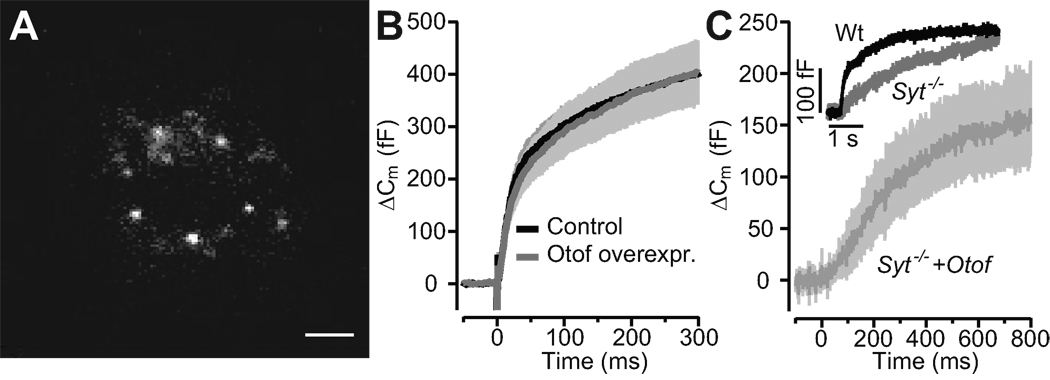
Figure 5. Overexpression of otoferlin in chromaffin cells does not change kinetics or amount of exocytosis and does not restore synchronous exocytosis in Syt1 deficient cells.
A, TIRF image of the footprint of a chromaffin cell, which had been transfected with a otoferlin-eGFP fusion construct. Individual fluorescent spots most probably represent otoferlin-eGFP-tagged chromaffin granules, scale bar: 1 µm.
B, average ΔCm in response to the first flash in control (n = 20; black) and otoferlin-expressing (n = 20; red) bovine chromaffin cells. The first flash was delivered 120–180 s after establishment of the whole-cell configuration.
C, exocytic responses of a wild-type non-transfected mouse chromaffin cell (black) and mean response of 5 Syt1 deficient chromaffin cells that had been transfected to express the otoferline-GFP fusion construct: lack of the fast component of the exocytic burst. Insert: representative exocytotic response from wild-type (black, postflash [Ca2+]i: 16.31 µM) and Syt1 knock-out (red, postflash [Ca2+]i: 15.5 µM).

