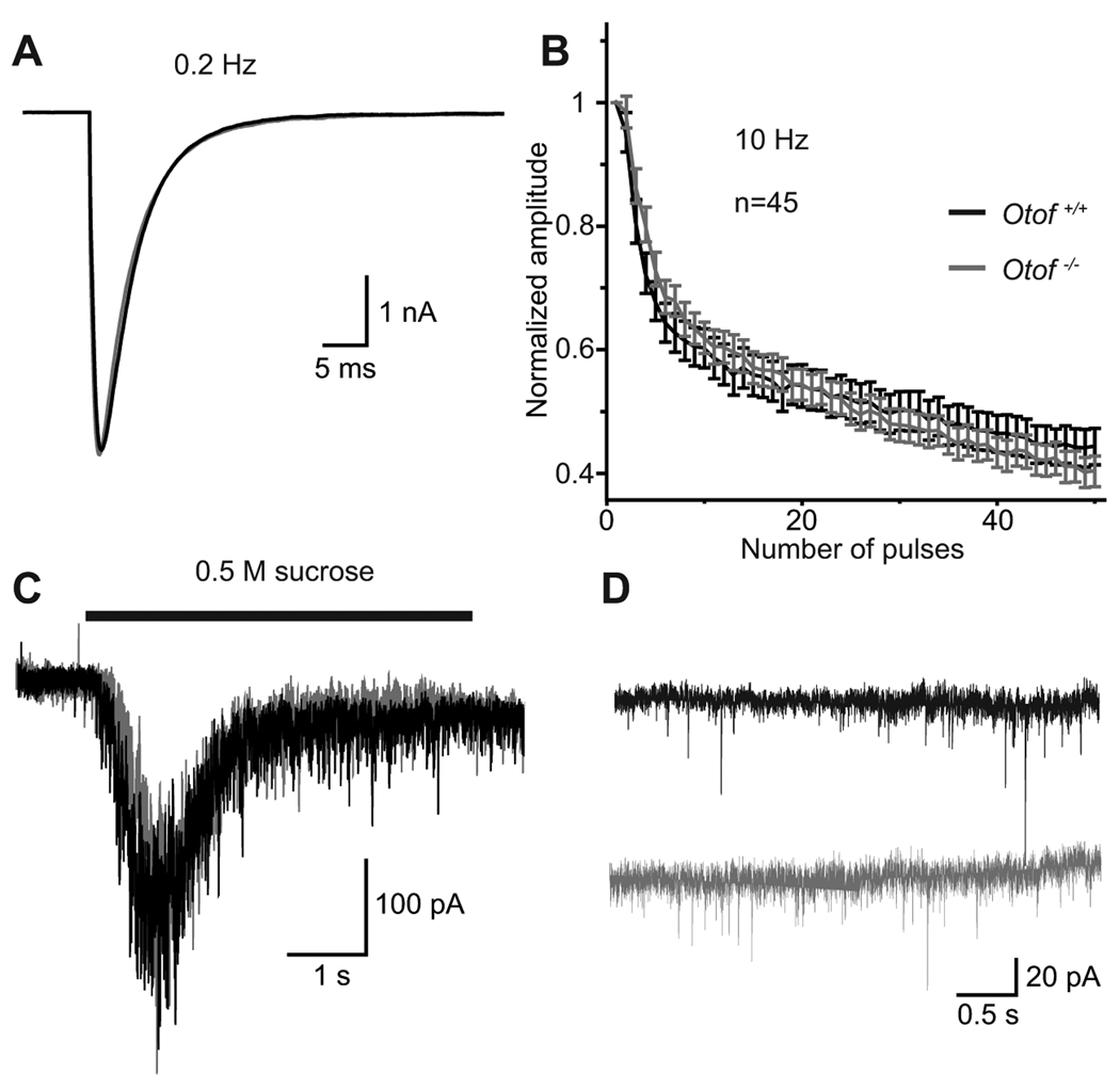
Figure 7. Exocytosis in Otof−/− hippocampal neurons.
A, representative EPSC traces from Otof+/+ (black) and Otof−/− (grey) autaptic hippocampal neurons at 10 to 15 DIV, elicited by depolarizations to 0 mV for 2 ms at 0.2 Hz.
B, mean normalized amplitudes of EPSCs during a train of action potentials at 10 Hz from Otof+/+ and Otof−/− hippocampal neurons.
C, representative traces showing the release of the readily releasable pool (RRP) induced by application of 0.5 M sucrose.
D, representative traces of miniature EPSC (mEPSCs) in the presence of 300 nM TTX at −70 mV. The frequency and the amplitude of mEPSCs was unchanged in Otof−/− neurons.

