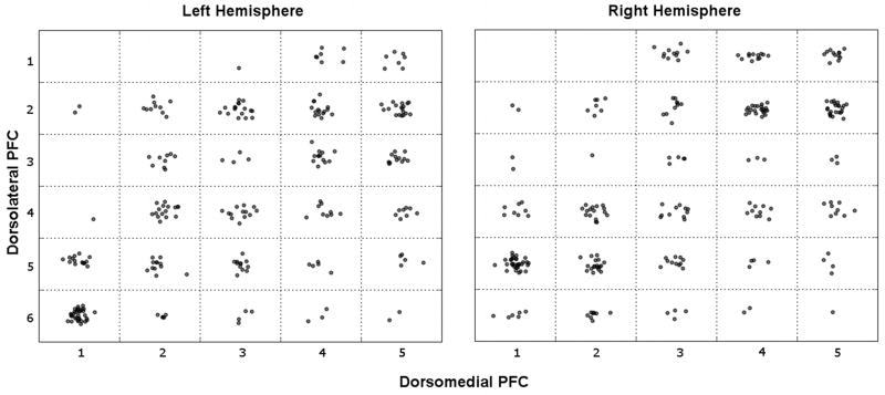
Figure 4. Individual participants' maximally correlated dmPFC-dlPFC pairs reveal parallel topography.
Each cell contains one dot for each participant whose maximal connectivity with a given dmPFC seed (x-axis) was to a given dlPFC seed (y-axis). Dots are spatially jittered within a cell for clarity. Even with this very conservative approach – which only considers the single maximal connection, without regard for the distribution – there is an increase in density along the diagonal.

