
Figure 1.
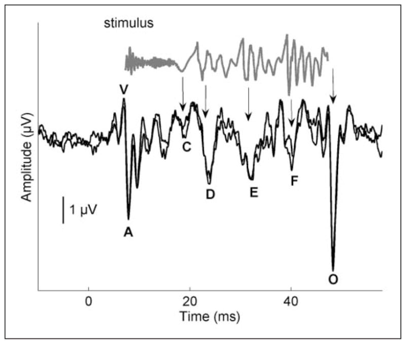
Time domain of a 40-ms stimulus /da/ (gray) and response (black)
Note. The stimulus evokes characteristic peaks in the response, labeled V, A, C, D, E, F, and O. The stimulus waveform has been shifted to account for neural lag and to allow visual alignment between peaks in the response and the stimulus. The arrows indicate where peaks in the stimulus correspond to peaks in the response. Two response waveforms of an individual participant are included to demonstrate replicability. Modified from Skoe and Kraus (2010).