
Figure 3.
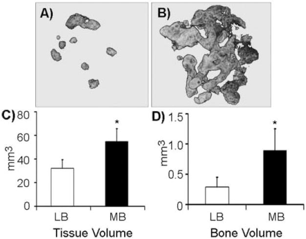
3D-reconstructed (A,B) microCT images of representative gelatin sponges seeded with (A) long-bone- or (B) mandible-derived marrow cells. (C) Tissue volume (TV) and (D) bone volume (BV) of long-bone (LB) vs. mandible (MB) marrow cell-seeded sponges, quantified by μCT (average of 8 individual transplants from 2 independent experiments). *p < 0.05; error bars represent standard error of the mean.