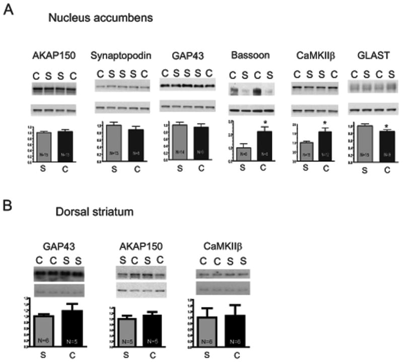
Figure 6. Western blot analysis of seven proteins identified by iTRAQ analysis as being significantly different between cocaine-trained and yoked saline subjects.

Equal amounts of PSD-enriched proteins (5-10 μg) were loaded per lane. Membranes were cut horizontally for simultaneous analysis of experimental protein (top) and LGN/AGS5 (bottom). Bands from chronic cocaine (C) and saline (S) administering animals are indicated. A, PSD subfractions prepared from the nucleus accumbens. B, PSD subfractions were identically prepared from dorsal striatum. Data are shown as percent change from yoked saline values. * p<0.05 comparing cocaine to saline using a Student's two-tailed t-test.
