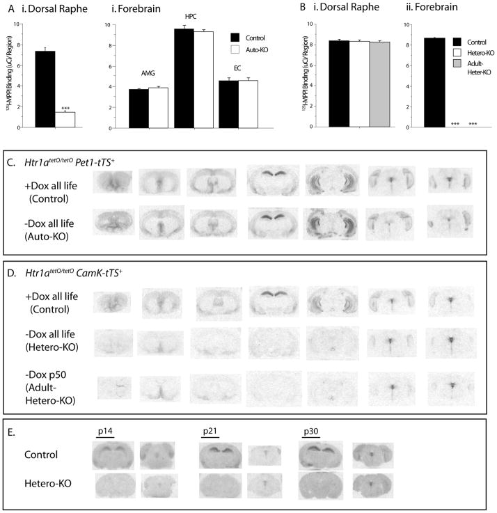
Figure 2.
(A) Quantitative 125I-labelled MPPI autoradiography for 5-HT1A receptors revealed a significant decrease in the dorsal raphe (i) of Auto-KO mice compared to their matched Controls. Conversely, no difference was detected in forebrain regions (ii), such as the ventral dentate gyrus of the hippocampus (HPC), the amygdala (AMG), and the entorhinal cortex (EC). (B) (i)No difference was detected in binding in the dorsal raphe of Hetero-KO or Adult-Hetero-KO mice compared to their matched controls. Conversely, binding was significantly decreased in dorsal CA1 of the hippocampus (HPC) in both groups compared to control (ii) (N=3–7/group). All values are mean ± SEM. ***p<0.0001. Matched autoradiograms showing detailed expression patterns of 5-HT1A receptors binding across the rostrocaudal extent of the brain in coronal section comparing (C) Auto-KO mice and their Controls; and (D) Hetero-KO and Adult-Hetero-KO mice and their Controls. Low levels of heteroreceptors remain in parts of the septum, olivary nucleus, and entorhinal cortex. (E) Receptor binding in the postnatal developmental period in Hetero-KO mice and their Controls. Matched sections are shown at the level of the dorsal hippocampus and the dorsal raphe nucleus. Forebrain receptor suppression is complete by P14.

