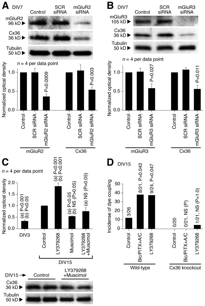
Figure 6.
Characterization of the mechanisms for developmental increase in neuronal gap junction coupling. Western blot (A–C) and dye coupling (D) tests were performed in neuronal cultures prepared from the rat (A–C) and mouse (D) hypothalamus. A, B, siRNA suppression (on DIV3-DIV7) of mGluR2 (A) and mGluR3 (B) decreases both the receptor and Cx36 protein levels. Representative images (above) and statistical data (below; paired Student’s t-test relative to control; mean ± SEM) are shown. Stainings were done sequentially on one membrane. SCR, scrambled siRNA. C, A combined activation (on DIV3-DIV15) of group II mGluRs and GABAARs (with LY379268 plus muscimol) does not affect significantly the developmental up-regulation of Cx36 expression. Statistical analysis: ANOVA with post hoc Tukey; (a), relative to DIV15 control; (b), relative to LY379268 plus muscimol; mean ± SEM. D, In wild-type cultures, dye coupling increases between DIV3 (not shown) and DIV15 (Control) and this increase is augmented by inactivation of GABAARs (Bic/PiTX+A/C) and by activation of group II mGluRs (LY379268). In Cx36 knockout cultures, neither the developmental nor the treatment-mediated increases occur. The number of dye-coupled primary-labeled neurons of the total number of primary-labeled neurons and statistical significance (Fisher’s exact probability test; relative to the corresponding control) on DIV15 are shown. Pi, P value cannot be calculated.

