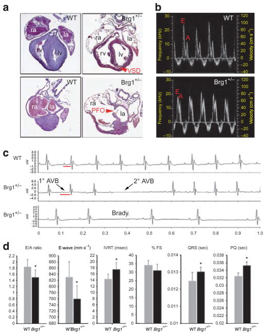
Figure 4. CHDs in Brg1 heterozygous null mice.
(a) Histology of postnatal day 0 WT (left panels) and Brg1 +/− hearts (right panels), showing dilated chambers, muscular ventricular septal defect (VSD) and patent foramen ovale (PFO) in Brg1 +/− hearts. Top and bottom panels are planes of section of the same heart at the level of the outflow tract (top panels) and at the level of the atrial septum (bottom panels). Original magnification: ×50. (b) Doppler waveforms of flow at the mitral valve of adult WT and Brg1 +/− mice, showing altered E and A wave amplitudes in Brg1 +/− mice. (c) ECG telemetry in WT and Brg1 +/− mice, showing prolonged PQ interval, sinus pause and second-degree atrioventricular block in Brg1 +/− mice. (d) Quantitation of selected parameter in WT (grey bars) and Brg1 +/− (black bars) mice. Units of measure are indicated in parentheses next to the graphed metric title. Data are mean ± s.d.; n = 5; *P < 0.05. la, left atrium; lv, left ventricle; ra, right atrium; rv, right ventricle.

