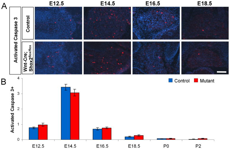
Figure 4. Apoptosis is normal in Wnt1-Cre; Shox2flox/flox DRG.
A. Representative images of immunostaining with anti-activated-caspase-3 in control and Shox2-deleted DRG at E12.5, E14.5, E16.5, and E18.5. B. Quantifications show no significant differences in the amount of apoptotic cells between control and Shox2-deleted DRG. Note that at E14.5, a time point of natural occurring cell death, there is an increase in activated-caspsase-3 positive cells in both control and mutant DRG. Error bars represent ±SEM. Scale bar = 100μm.

