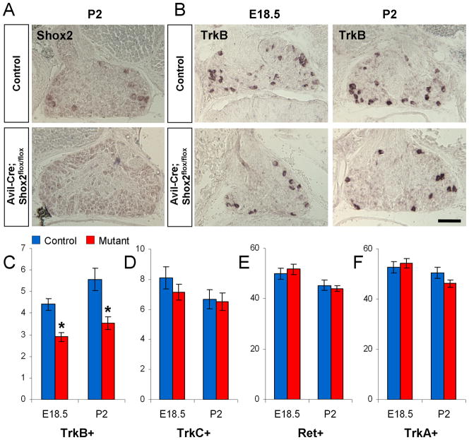
Figure 6. Mild reduction in the number of TrkB-expressing cells in AvilCre/+; Shox2flox/flox mice.
A. In situ hybridization confirms the loss of Shox2 expression in the DRG from AvilCre/+; Shox2flox/flox mice. B. Representative images of TrkB expression in control and AvilCre/+; Shox2flox/flox mice at E18.5 and P2. C–F. Quantifications of the numbers of TrkB (C), TrkC (D), Ret (E), and TrkA (F) expressing DRG neurons per unit area in control and AvilCre/+; Shox2flox/flox mice at E18.5 and P2. *p<0.001. Error bars represent ±SEM. Scale bar = 100μm.

