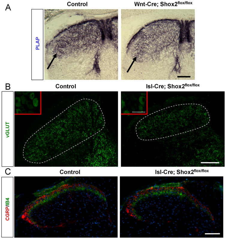
Figure 8. Reduced mechanosensory central innervation in the spinal cord in Shox2-deleted mice.
A. Representative images of sensory afferent projections in the spinal cord from control (Wnt1-Cre; Shox2flox/+;AvilPLAP/+) and Shox2-deleted mice (Wnt1-Cre; Shox2flox/flox;AvilPLAP/+) as revealed by PLAP staining at P0. Arrows point to a densely stained band in layer III/IV in control that is only moderately stained in the mutant. B. Representative images of immunofluorescence staining with anti-vGluT1 in the spinal cord of control (Isl1-Cre; Shox2flox/+) and Shox2-deleted (Isl1-Cre; Shox2flox/flox) mice. Inset shows the anti v-GluT1 immunofluorescence signal in the DRG of control and Shox2-deleted mice. C. Representative images of immunofluorescence staining with anti-CGRP (red) and anti-IB4 (green) in the spinal cord of control and Shox2-deleted mice. Blue is DAPI. Scale bar in A and C is 100μm. Scale bar in B is 50μm.

