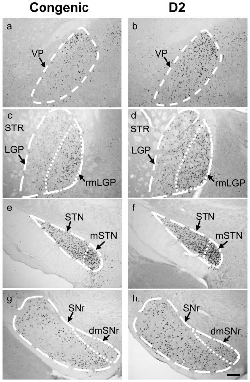
Figure 2.
Photomicrographs of c-Fos expression in representative brain regions in ethanol withdrawn Alcw2 congenic and background strain (D2) mice. Congenic mice showed significantly fewer c-Fos immunoreactive cells within the ventral pallidum (VP) (a), rostromedial subregion of the lateral globus pallidas (rmLGP) (c), subthalamic nucleus (STN) (e), and substantia nigra pars reticulata (SNr) (g) than D2 background strain mice (b,d,f,h), respectively. Subregions associated with limbic function [e.g., rmLGP, medial STN (mSTN), and dorsomedial SNr (dmSNr)] are delineated in panels c–h. The medial subregions of the rostral LGP (c,d) and STN (e,f) were more intensively activated than the lateral subregions. In rostral SNr, the limbic subregion (i.e., dmSNr) showed increased neuronal activity associated with ethanol withdrawal in both strains, but to a lesser degree in congenic vs. background strain mice (g,h). Additional abbreviations: STR, striatum. Scale bar = 100 μm.

