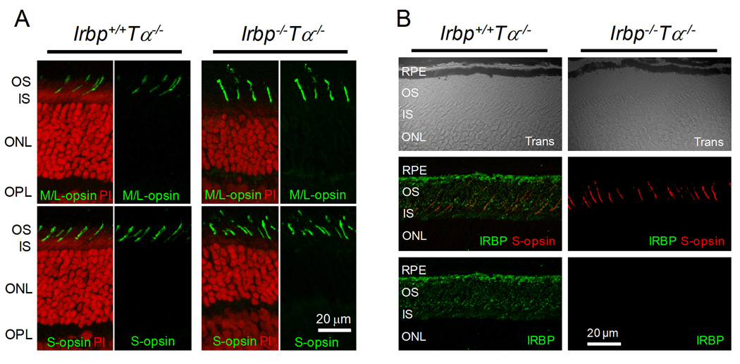
Figure 2.
Cone opsin localization in 2-month-old Irbp−/−Tα−/− mice. A, Both M/L- and S-opsins are localized in cone outer segments of Irbp+/+Tα−/− and Irbp−/−Tα−/− mice. Cross section images of the retina (central-ventral location to the optic nerve head) are shown. OS: outer segments, IS: inner segments, ONL: outer nuclear layer, OPL: outer plexiform layer. Scale bar, 20 µm. Cell nuclei were stained with propidium iodide (PI). B, Immunostaining of Irbp+/+Tα−/− and Irbp−/−Tα−/− retinas (central-ventral retina location) with anti-IRBP antibody. Trans: confocal images in transmitted light. Scale bar, 20 µm.

