Figure 5.
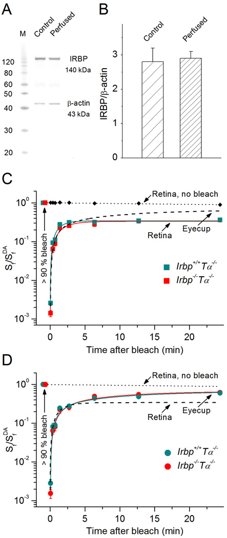
M/L-cone dark adaptation in Irbp−/−Tα−/− mice. A, Representative Western blot of Irbp+/+Tα−/− eyecup before and after 30 min perfusion to determine levels of endogenous IRBP. β-actin served as a loading control. Both IRBP and β-actin amounts were within the linear range of the Western blot sensitivity (not shown). B, Quantification of the data shown in (A) (n = 4). Values are means ± SEM.C, D, Recovery of cone a-wave flash sensitivity (Sf) in mouse retinas (C) and eyecups (D) after bleaching > 90% of cone pigment at time 0 (with 505 nm LED light). Retinas: Irbp+/+Tα−/− (n = 9), Irbp−/−Tα−/− (n = 7). Eyecups: Irbp+/+Tα−/− (n = 11), Irbp−/−Tα−/− (n = 8). Retina data (C) were fitted with single-exponential functions that yielded time constants of ~ 1.2 min and ~ 1.7 min, respectively. Eyecup data (D) were then fitted with double-exponential functions, and their initial rapid phase was fitted using the same time constants as determined for retinas (fixed parameter). R2 = 0.98. The slow RPE-driven cone recovery time constants for Irbp+/+Tα−/− and Irbp−/−Tα−/− animals were ~ 13 min and ~ 10 min, correspondingly. Values are means ± SEM. SfDA indicates sensitivity of dark-adapted preparations. Dashed lines show the cone a-wave flash sensitivity recovery (averaged data from the two mouse lines) in eyecups (C) and retinas (D), for comparison. Small black symbols connected with the dotted line (C) or the dotted line alone (D) show the stable flash sensitivity of dark-adapted retina during the 30 min recording session (a combined data from Irbp+/+Tα−/− and Irbp−/−Tα−/− retinas, n = 8).

