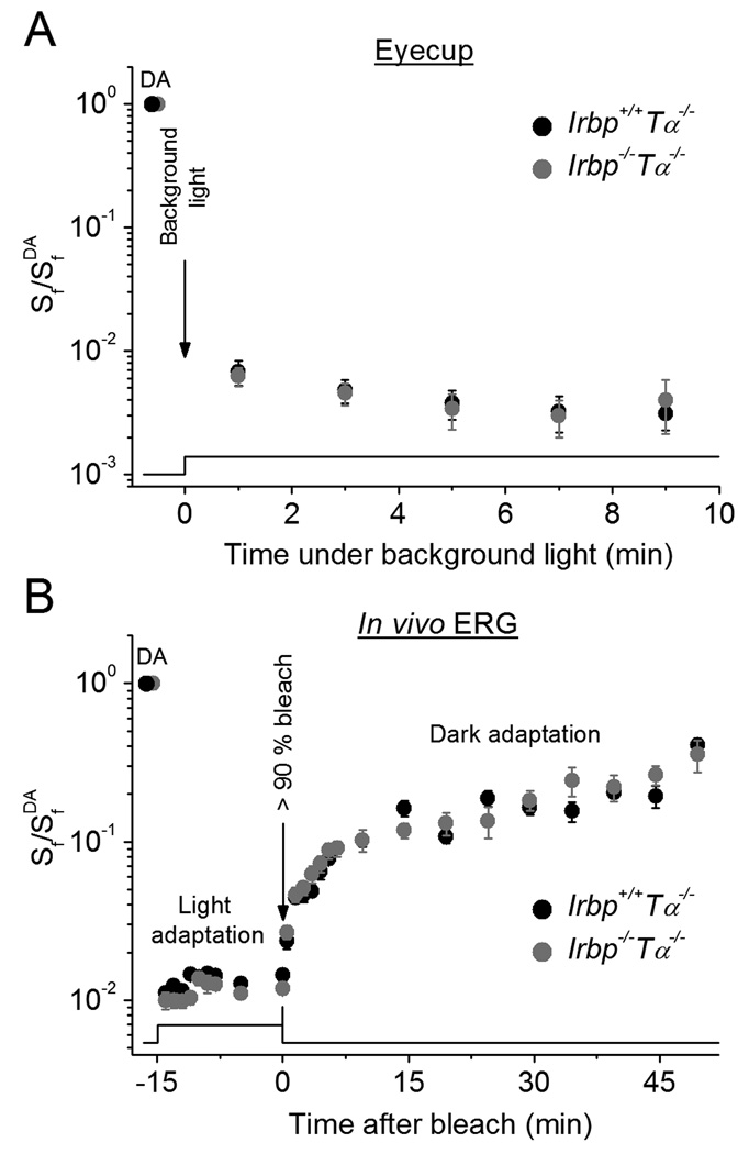
Figure 6.
M/L-cone light adaptation in Irbp−/−Tα−/− mice. A, Change of eyecup cone a-wave flash sensitivity (Sf) after applying constant background 505 nm LED light (2.9 × 10⁶ photons µ m−2 s−1).Irbp+/+Tα−/− (n = 9), Irbp−/−Tα−/− (n = 9). Values are means ± SEM (smaller than symbol size for most data points). SfDA indicates flash sensitivity of dark-adapted eyecups.B, Change of photopic ERG b-wave flash sensitivity (Sf) after applying white Ganzfeld background light (514 cd m−2, 15 min) and its subsequent recovery (in darkness) after bleaching > 90% of cone pigment. Live animals: Irbp+/+Tα−/− (n = 5 mice), Irbp−/−Tα−/− (n = 5 mice). Flash sensitivity was normalized to its dark-adapted value (SfDA). Bleaching was achieved by 30 s illumination with 520 nm LED light at time 0. Values are means ± SEM. The timecourse of light stimulation in (A, B) is shown on the bottom.

