Figure 1.
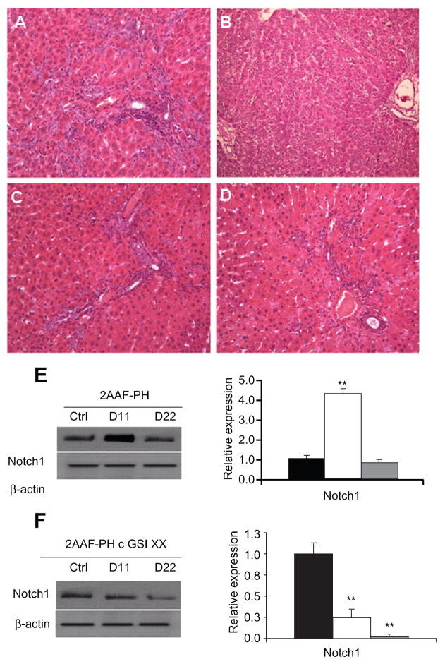
Hepatic oval cell activation and detection of notch expression. A) Representative hematoxylin and eosin staining of liver section taken from animals on the 2AAF-PH protocol alone on day 11 post-PH. B) Representative hematoxylin and eosin staining of liver section taken from animals on the 2AAF-PH protocol alone on day 22 post-PH. C) Representative hematoxylin and eosin staining of day 11 liver section taken from animals on the 2AAF-PH protocol and treated with GSI XX. D) Representative hematoxylin and eosin staining of day 22 liver section taken from animals on the 2AAF-PH protocol and treated with GSI XX. in A, C, and D, “streaming” oval cells (black arrows) can be seen between portal triads (periportal regions); this phenomenon is absent in B, where the oval cells have differentiated into mature lineages by day 22 post-PH. in C, the white arrow indicates cells part of the immune infiltrate (small, dark, punctate cells), which have mostly disappeared by day 22 (D) because the vehicle/inhibitor have been processed out of the liver. E) Left: Western blot analysis performed on protein isolated from liver taken at day 11 and day 22 post-PH alone with an antibody specific for the NICD, or cleaved (activated) portion of the Notch1 receptor. Right: semiquantitative analysis of Notch1 protein in the 2AAF-PH alone group for control, day 11 post-PH, and day 22 post-PH samples. Expression was normalized to β-actin and significance calculated compared with control animals. *P < 0.01. F) Left: Western blot analysis performed on protein isolated from liver taken at day 11 and day 22 post-2AAF-PH with an antibody specific for NICD, or cleaved (activated) portion of the Notch1 receptor. Right: semiquantitative analysis of Notch1 protein in control, day 11 post-PH, and day 22 post-2AAF-PH livers from animals treated with GSI XX. Expression was normalized to β-actin and significance calculated compared with control animals. *P < 0.01. A–D 200×.
Abbreviations: NiCD, Notch intracellular cytoplasmic domain; GSI XX, γ-secretase inhibitor; PH, partial hepatectomy; 2AAF-PH, 2-acetylaminofluorine implantation followed by 70% surgical resection of the liver.

