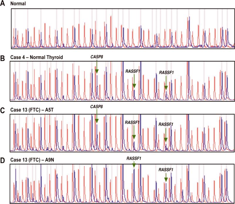
Figure 2.
A: Normal control DNA sample with 41 individual peaks (red) in the absence of HhaI and 15 separate peaks (blue) in the presence of HhaI. B: Normal thyroid sample with methylation of CASP8 and RASSF1. C: Follicular thyroid cancer (Case 13 - tumor block) with methylation of CASP8 and RASSF1. D: Follicular thyroid cancer (Case 13 - normal block) with methylation of RASSF1. (FTC – follicular thyroid cancer, A5T – block A5 tumor, A9N – block A9 normal)

