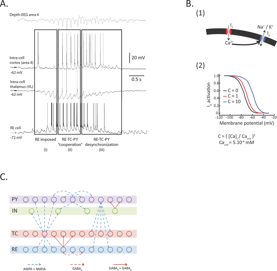
Fig. 1. Basic spindle properties and model topology.
A. Field potential and simultaneous recordings from cortical and thalamocortical. A recording from reticular thalamic neuron was obtained in a different experiment. (Modified from Timofeev & Bazhenov, 2005).
B. (1) [Ca2+]i-dependent shift in the activation curve of the hyperpolarization-activated h-type current, Ih. T-type current, IT, allows Ca2+ ions to enter the cell, which bind to the mixed Na+/K+ channel Ih and modify its voltage-dependent properties. (2) Binding of Ca2+ ions to Ih channels shifts the voltage dependence of the current towards positive membrane potentials.
C. Model topology and structure of the thalamocortical network distributed into four different layers. The thalamocortical relay (TC) neurons and the thalamic reticular (RE) neurons formed two of the layers, strongly interacting with each other. The two cortical layers contained pyramidal excitatory (PY) neurons and inhibitory interneurons (IN). Connections between layers were stochastically distributed. Heterogeneity in the same layer was achieved through a random distribution of parameters for intrinsic properties (see Methods for details).

