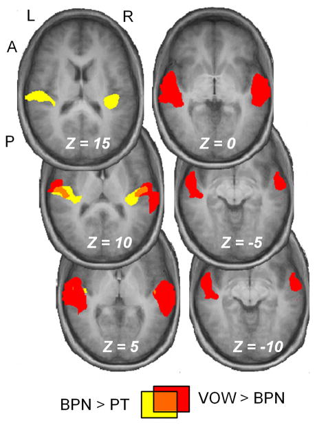
Figure 2. A progression from simple to complex selectivity in the antero-lateral direction.
A BPN > PT contrast (yellow) yielded activation largely restricted to areas adjacent to the medial and lateral extents of Heschl’s gyrus in each hemisphere. The VOW > BPN contrast yielded significant activation in antero-lateral aspects of the superior temporal gyrus. The two contrasts share little overlap (orange). Thresholds were P < 0.05 for both contrasts, and the sizes and Talairach coordinates of the resulting clusters are reported in Table 2.

