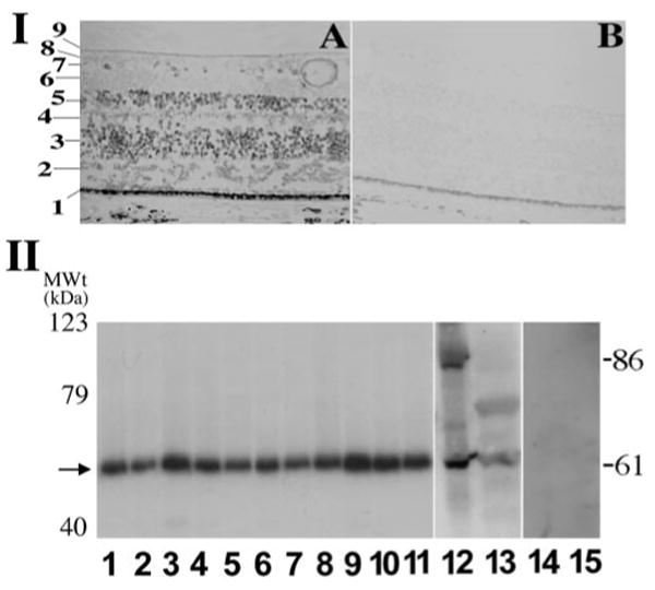
Figure 1.

Detection of human PRPF31 gene expression by in situ hybridization and by Western blotting assays. I, Expression of PRPF31 gene in the retina, as detected by in situ hybridization. Cells in different layers of the retina express PRPF31, as detected by the PRPF31 antisense probe (A). The sense control probe does not show detectable signal (B). Distinct histologic layers of the retina are marked as follows: 1, retinal pigment epithelium; 2, photoreceptor cell layer; 3, outer nuclear layer, nuclei of photoreceptor cells; 4, outer plexiform layer; 5, inner nuclear layer; 6, inner plexiform layer; 7, ganglion cell layer; 8, retinal nerve fiber layer; 9, internal limiting membrane. II, Western blotting results showing a prominent band of 61 kDa in a range of adult murine tissues (lanes 1–9; 1, brain; 2, heart; 3, kidney; 4, spleen; 5, lung; 6, thymus; 7, muscle; 8, lymph node; 9, retina), in postnatal retina (from P2 mice, lane 10), in embryonic retina (from embryonic day 18 mice, lane 11), and in human HEK cells as well as stable HEK cells expressing a PREF31–GFP fusion protein (lanes 13 and 12, respectively). Protein concentrations of the cell lysates were measured, and equal amounts were loaded in each lane. These bands represent specific PRPF31 signals (61 kDa for the endogenous protein and 86 kDa for PRPF31–GFP fusion protein), because they are not detectable when preimmune antibody preparation was used (lanes 14 and 15). There is no significant change in the gel mobility of PRPF31 protein detected in different tissues. MWt, Molecular weight.
