Fig. 5.
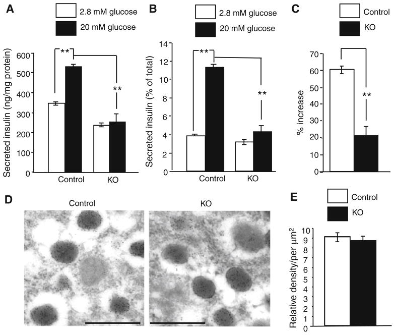
Reduced insulin release from pancreatic islets isolated from Ins2-Hap1−/− (KO) mice. a–c Secreted insulin is presented as ng/mg protein in (a) the percentage of total insulin content (b), and the increased percentage of the basal level in (c). Control mice (Ins2-Hap1 ±) mice were used for comparison (n = 6, **p <0.01). d, e Immunogold electron microscopic examination of the pancreatic islets from control and KO mice. The morphology (d) and the density (e) of insulin-containing granules are not significantly different between control and KO mice. Scale bars 1.0 μm

