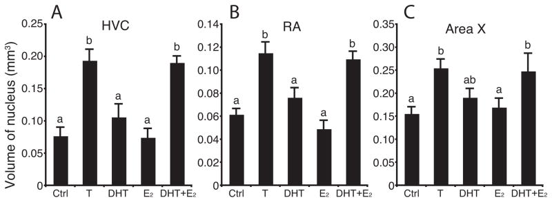
Figure 2.
Testosterone (T) or a combination of its androgenic (DHT) and estrogenic (E2) metabolites increased the volume of HVC (left), RA (middle) and Area X (right) in female canaries. Data for each nucleus were analyzed by one-way ANOVAs followed by Fisher’s least significant difference (LSD) post hoc tests (letters above the bars). Bars with different letters are significantly different for P < 0.01 in HVC and for P < 0.05 in RA and Area X.

