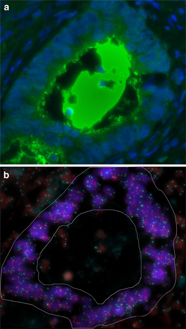
Fig. 2.
IHC and FISH image of the same tumour gland. a: Immunofluorescence of a CD133 positive tumour gland. The CD133 protein expression is localized to the glandular-luminal surface and intraluminal of the tumour. b: FISH for ZNF217 (Spectrum Orange) and centromere 6 (Spectrum Aqua) in the same gland as shown in a. Only the tumour gland (the area within the demarcation) was selected for evaluation, the normal surrounding cells as well as apoptotic cells in the lumen were excluded. This case was amplified for ZNF217 showing a ratio (ZNF217/CEP6) of 1.4

