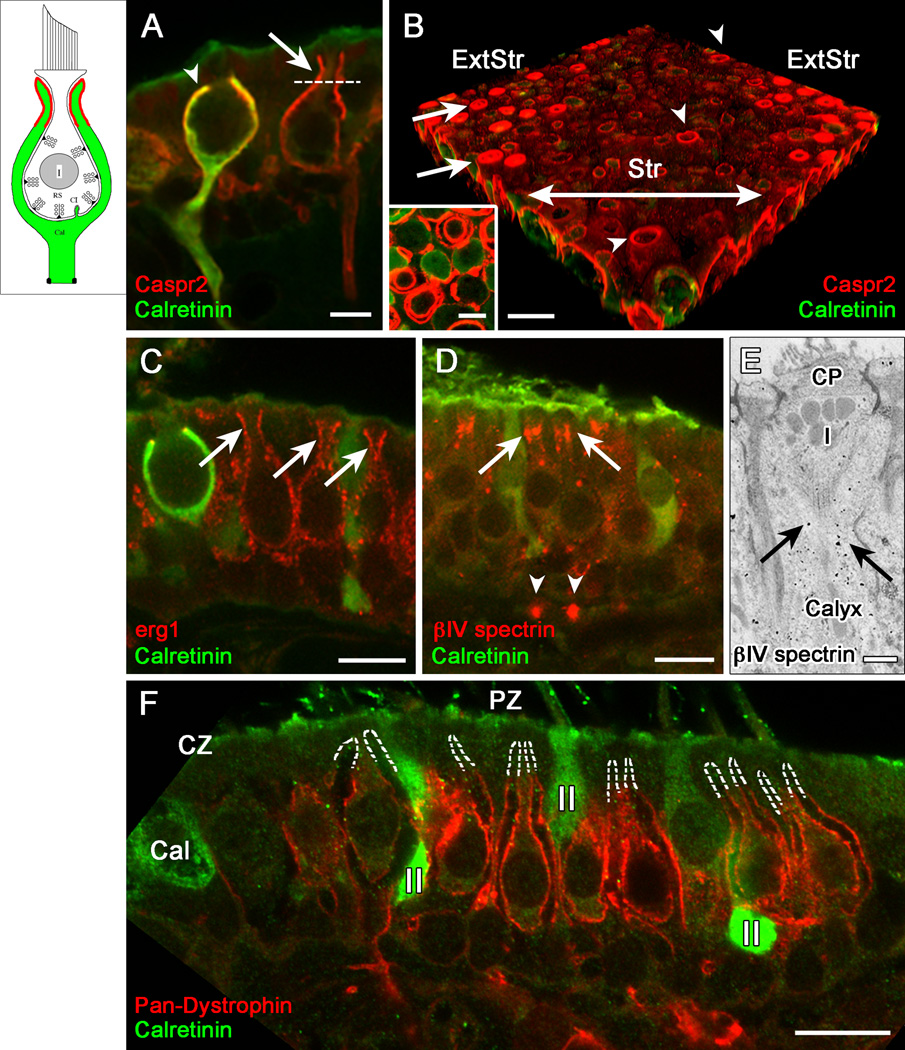
Figure 2.
Domain 2, the apical part of the calyx, was selectively labeled by antibodies to the cell adhesion molecule, Caspr2, and to erg KV subunits, and lacked several proteins found in Domain 3. (A, B) Caspr2 antibody (red) outlines Domain 2: the apical part of the calyx (above the dashed line in A). These central calyces belong to a calyx-only (calretinin-positive) and a dimorphic (calretinin-negative) afferent. Domain 2 extends higher in dimorphic afferents (arrows, in A and B) than in most calyx-only afferents (arrowhead, in A), as shown in confocal transverse sections (A) and 3D reconstructions of whole-mount utricular maculae (B). In B, the striola (Str) had much less Caspr2 staining (red) than did the extrastriolar region (ExtStr). Arrowheads in B indicate Caspr2 label of three calyces of striolar dimorphic afferents, which extended to the apical surface of the epithelium. A transverse optical section of Domain 2 (inset), taken above the level of the dotted line in A, shows concentric circles of Caspr2 label, demonstrating that it is present on both the inner and outer calyx surfaces. (C) Domain 2 was labeled by antibodies to erg1 and erg2, but not by any of the other KV antibodies we used (KCNQ2, 3, 4, 5; KV1.1, 1.2). Erg1 antibody labeled Domain 2 in calretinin-negative dimorphic afferents (arrows) but not in calyx-only afferents, as shown by the calretinin-positive (green) calyx on the left. Erg2 (not shown) had a similar distribution. (D, E) Immunoreactivity for βIV spectrin, which is usually found at nodes (arrowheads in D point to heminodes on the afferent fibers) and axonal initial segments, is also found in Domain 2 (arrows), shown by both confocal immunohistochemistry (D) and EM immuno-gold localization (E). Note the gold particles (E, arrows) in the calyx (Cal) surrounding the upper part of a type I (I) hair cell below the cuticular plate (CP). (F) Domain 2 is also defined by the absence of several Domain 3 proteins, including dystrophin (red); dashed white lines outline the unlabeled Domain 2 in several calyces of peripheral-zone (PZ) dimorphic afferents. Dimorphic afferents are calretinin-negative, unlike central-zone (CZ) calyx-only afferents (green, Cal) and peripheral-zone (PZ) type II hair cells (II). Scale bars: A, B inset, 5 µm; B–D, F, 10 µm; E, 500 nm.

