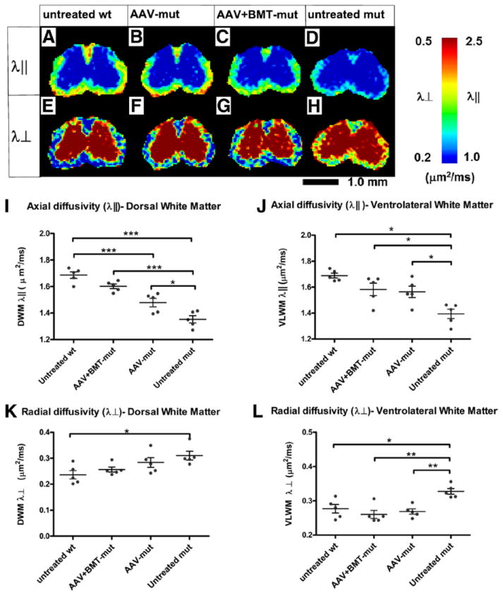
Figure 5.

Diffusion tensor imaging. Heat maps of axial diffusivity (λ∥; A–D) and radial diffusivity (λ⊥; E–H) in the spinal cord obtained by DTI. In the DWM (I), there is a significant decrease in the axial diffusion in the untreated mut group compared with the untreated wt group. The AAV-mut and AAV+BMT-mut groups show an increase in axial diffusion compared with the untreated mut group. In the VLWM (J), the axial diffusivity of the untreated mut is significantly decreased compared with the untreated wt and the treated groups. Radial diffusivity in the DWM (K) is significantly increased in the untreated mut compared with the untreated wt. The treated groups are intermediate between the untreated wt and untreated mut groups. In the VLWM (L), there is a significant increase in the radial diffusivity in the untreated mut compared with untreated wt. There is no significant difference between the untreated wt and the AAV-mut and AAV+BMT-mut groups. The horizontal bars represent the means, and the error bars represent SEM. *p < 0.05; **p < 0.01; ***p < 0.001; ns, not significant.
