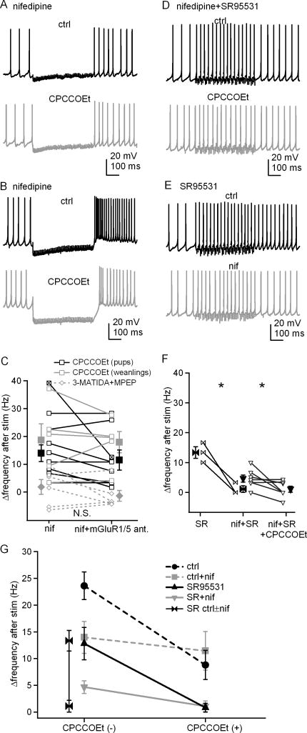
Figure 7. Prolonged rebound firing is reduced by blockade of L-type Ca channels.
A, Action potentials in a cerebellar nuclear cell, with a 500-ms, 100-Hz stimulus train before and after application of CPCCOEt in nifedipine. Fast excitatory transmission was blocked by DNQX and CPP. B, Action potentials recorded from the one cell showing rebound burst firing under the same conditions as in A, ± CPCCOEt. C, Summary of firing rate changes after stimulus trains in nifedipine ± CPCCOEt or nifedipine ± 3-MATIDA and MPEP. Open symbols represent individual cells and solid symbols indicate mean ± SEM. The x-box symbol indicates the bursting cell in B, and was excluded from the statistical analysis. Black squares, data from pups (P13–P15) with CPCCOEt; grey squares, data from weanlings (P22–P28) with CPCCOEt; grey diamonds, data from pups with 3-MATIDA and MPEP. D, Action potentials from another cell under the same conditions as in A, except with inhibitory input blocked by SR95531. E, Action potentials from a cell with stimulus trains applied before and after exposure to nifedipine without CPCCOEt, with inhibitory and fast excitatory transmission blocked. F, Summary of firing rate increases ± nifedipine and/or CPCCOEt, with inhibitory input blocked. G, Summary of firing rate changes for all conditions tested.

