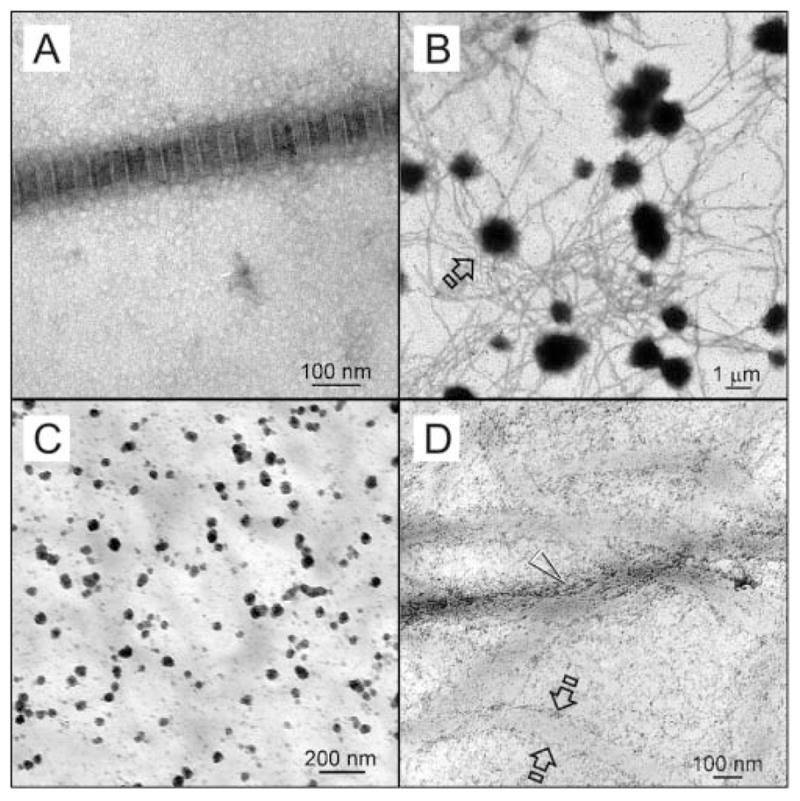
Figure 2.

TEM images of collagen fibrils before mineralization and those obtained from controls. (A) Negative-stained, cross-linked reconstituted collagen fibril with approximately 76 nm periodicity. (B) Unstained image of the sequestration analog negative control. Electron-dense microspheres (open arrow; ca. 0.5–2 μm) were formed around unmineralized collagen fibrils after they were phosphorylated with HPA-Na3P3O9. However, no intrafibrillar mineralization was observed after 24 hrs. (C) Unstained image of the matrix phosphoprotein analog negative control retrieved after 4 hrs, showing formation of electron-dense nanospheres (arrow; ca. 20–50 nm) in the vicinity of the unmineralized, non-phosphorylated collagen matrix when polyacrylic acid was included as an ACP-stabilization analog in the mineralization medium. (D) Unstained image of the matrix phosphoprotein analog negative control retrieved after 24 hrs. Minerals were formed along fibril surfaces (between open arrows). Intrafibrillar mineralization was seen as electron-dense microfibrillar strands, but no banding could be identified.
