Figure 4.

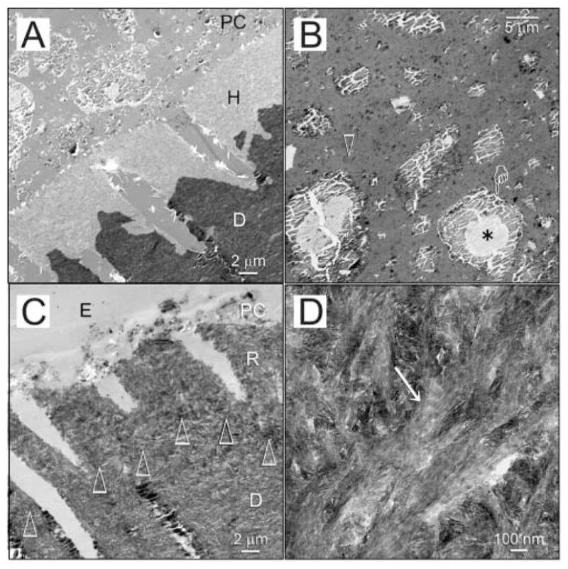
Biomimetic remineralization of acid-etched dentin with sodium trimetaphosphate. (A) Unstained image of acid-etched dentin (AE) phosphorylated with HPA-Na3P3O9 for 5 min and capped with a Portland-cement-containing composite (PC). No remineralization was seen after 1 mo of immersion in the sequestration analog-containing mineralization medium. D: mineralized dentin. (B) The composite contains set Portland cement and silanized silica (open arrow) within its resin matrix. The Portland cement particles contain a cement core (asterisk) surrounded by calcium silicate hydrate (pointer). (C) Unstained image of the completely remineralized layer (R) of acid-etched dentin after 3–4 mos of biomimetic remineralization. The original demineralization front is demarcated by open arrowheads. The composite (PC) had dislodged, leaving a thin layer embedded by laboratory epoxy resin (E). D: mineralized dentin. (D) Intrafibrillar mineralization (open arrowhead) within the remineralized layer.
