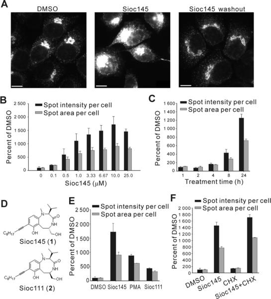
Figure 1.
Sioc145 enhances the signal of p58-YFP in H4 cells. (A) H4-p58-YFP cells were treated with DMSO or 5 μM Sioc145 for 24 h or then exchanged with fresh medium without Sioc145 for an additional 24 h. Images were analyzed for spots' intensity and area per cell. Scale bar, 10 μm. (B) Dose-response of Sioc145 in H4-p58-YFP cells treated for 24 h. (C) Time course response of 5 μM Sioc145 in H4-p58-YFP cells. (D) Chemical structures of Sioc145 and Sioc111. (E) p58-YFP signal in cells treated with DMSO, 5 μM Sioc145, 5 μM Sioc111 or 100 ng/ml PMA. (F) p58-YFP signal in cells treated with DMSO, 5 μM Sioc145, 10 μg/ml CHX alone or CHX together with 5 μM Sioc145 for 24 h. All graphics represent means ± SD obtained from three independent experiments.

