Figure 7.
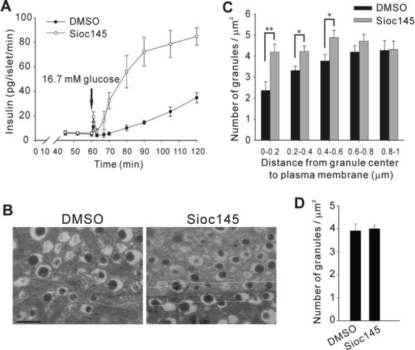
Characterization of secretion kinetics and insulin granule distribution in Sioc145-treated rat pancreatic islets. (A) Rat pancreatic islets pretreated with 5 μM Sioc145 or DMSO for 30 min were perifused with 16.7 mM glucose buffer for 1 h, and insulin secretion was measured at indicated time points (see Materials and Methods). (B) Islets were treated with DMSO or 5 μM Sioc145 for 30 min, followed by a 60 min equilibration in 2.8 mM glucose buffer. Representative electron micrographs of islet β-cell sections are shown. Scale bar, 0.5 μm. White lines indicate a distance of 0.2 μm from the plasma membrane. (C) Density of insulin granules located in 0.2 μm concentric shells within the first 1 μm area from the plasma membrane. Granules located in the single section were categorized according to their distance from the granule center to the plasma membrane. (D) The average density of granules in cytoplasm. The area of cytoplasm is calculated as the cell area minus the nuclear area. Error bars represent SEM from three independent experiments (A) or 10 individual β-cells derived from four rats (C, D). *P < 0.05; **P < 0.01.

