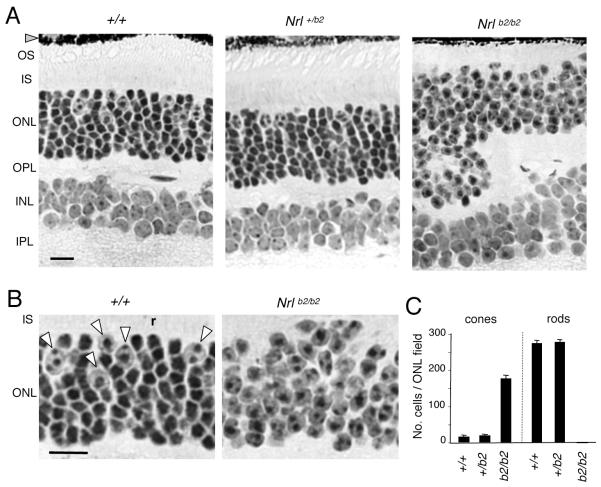
Figure 2. Retinal histology in Nrl+/b2 and Nrlb2/b2 mice.
A, Retinal histology in 3 month old mice. The outer nuclear layer (ONL) in Nrl+/b2 mice contains cones (large nuclei with dispersed chromatin) and rods (smaller, denser nuclei) in the same proportions as +/+ mice. Nrlb2/b2 mice like Nrl−/− mice lack rods, have excess cones and also display ONL folding and shortened outer segments (OS). INL, inner nuclear layer, IPL, inner plexiform layer, IS, inner segments, OPL, outer plexiform layer. Scale bar, 20 μm (same in B).
B, Higher magnification from images in panel A indicating cone nuclei (arrowheads) and a representative rod nucleus (r) in a +/+ mouse. Nrlb2/b2 mice possess only cone-like nuclei.
C, Counts (mean ± SD) of cone and rod nuclei in ONL fields, determined on 3 μm thick plastic sections like those in panel A. Fields avoided grossly folded areas in Nrlb2/b2 mice.

