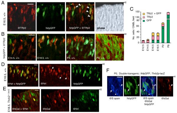
Figure 6. Transient co-expression of TRβ2 and Nrl in photoreceptor precursors.
A, Double fluorescence analysis of cells positive for TRβ2 (@TRβ2 antibody, red), NrlpGFP (transgene, green), or both @TRβ2 and NrlpGFP (yellow or orange, arrows in merged image) in the outer neuroblastic layer of +/+ mouse retina at E16.5. Right panel, phase contrast image of the same field. Confocal microscope images represent a single 1 - 1.5 μm z-plane obtained from 10 μm thick mid-retinal cryosections (same in panels B, C, D, E). For orientation, the grey arrowhead indicates location of retinal pigmented epithelium. Scale bar, 14 μm.
B, Double fluorescence analysis of cells positive for TRβ2 and NrlpGFP in +/+ mouse retina during development at E14.5, E18.5, P2 and P8. Doubly-positive cells (yellow or orange) were most evident at E16.5 - E18.5.
C, Counts of cells positive for TRβ2, GFP or both TRβ2 and GFP, determined on single z-plane confocal images from experiments shown in A and B. Counts shown were determined in mid-retinal fields. Counts in superior and inferior fields gave similar results.
D, Verification of NrlpGFP transgene as a marker for cells expressing endogenous Nrl using an antibody against NRL protein (@Nrl) and direct fluorescence for NrlpGFP in +/+ embryos at E18.5. The @Nrl+ population included almost all GFP+ cells (yellowish and orange cells, merged image in left panel) and a few @Nrl+ cells that were negative for GFP (white arrows, middle panel). Scale bar, 10 μm (same in D).
E, Independent identification of cells that co-express TRβ2 (@bGal) and endogenous NRL protein (@Nrl) (yellow or orange cells in merged image, left panel). Analysis was performed on E18.5 embryos homozygous for a targeted insertion of lacZ in the TRβ2-specific exon of the Thrb gene.
F, Immunofluorescence analysis for co-expression of S opsin (@S opsin antibody, blue), NrlpGFP (direct fluorescence, green) and Thrb2p-lacZ transgenes (@bGal antibody, red, indicator for TRβ2) in +/+ mice at P0. Arrow, S opsin/GFP doubly-positive cell; arrowhead, S opsin/GFP/bGal triply-positive cell; asterisk, S opsin-positive cell with no detectable GFP or bGal.

