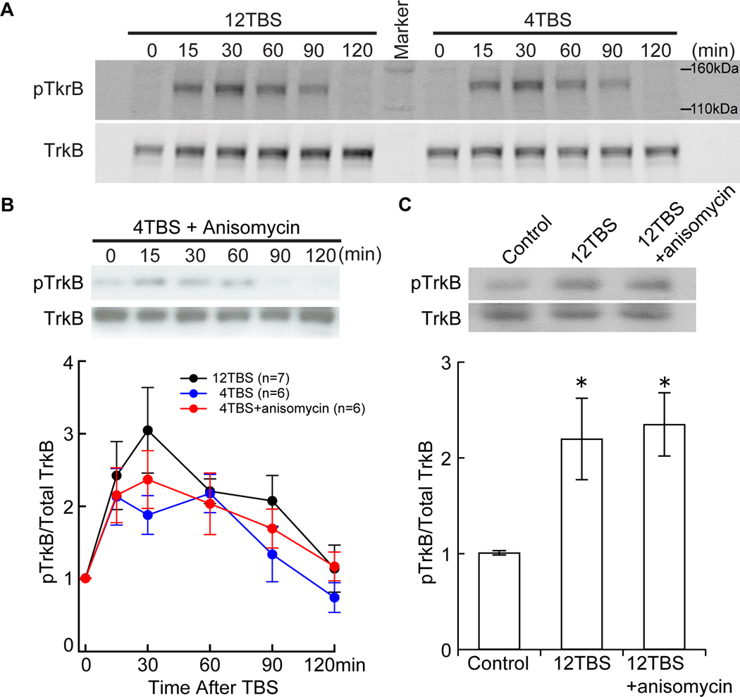
Figure 1. Transient and protein synthesis independent activation of TrkB by TBS.
CA1 areas were stimulated by stimulating Schaffer collaterals with 12TBS or 4TBS, dissected out quickly at different time points after TBS, and processed for Western blotting. For each individual time point, the ratio of pTrkB signal to total TrkB signal was normalized against the control condition from the same experiment. Three mice were used for each experiment with multiple time points, and each time point contains CA1 from 4 hippocampal slices. The same experiment was repeated at least 6 times (n = 6) in (A) and (B), each with new batch of animals.
(A) Examples of TrkB activation in hippocampal slices induced by 12TBS and 4TBS. No obvious difference was found in the pattern of TrkB activation.
(B) Quantification of time courses of TrkB activation induced by 4TBS with and without the protein synthesis inhibitor anisomycin (40 µM). The pTrkB time course induced by 12TBS is included for comparison.
(C) Protein synthesis independence of TrkB activation measured 1 hour after 12TBS. Both example (top) and quantification (bottom, n = 4) are shown. Inhibition of protein synthesis by anisomycin (40 µM) does not block the TBS-induced increase in pTrkB (*, P <0.05, one-way ANOVA; no difference was found between “12TBS” and “12TBS+anisomycin” groups).

