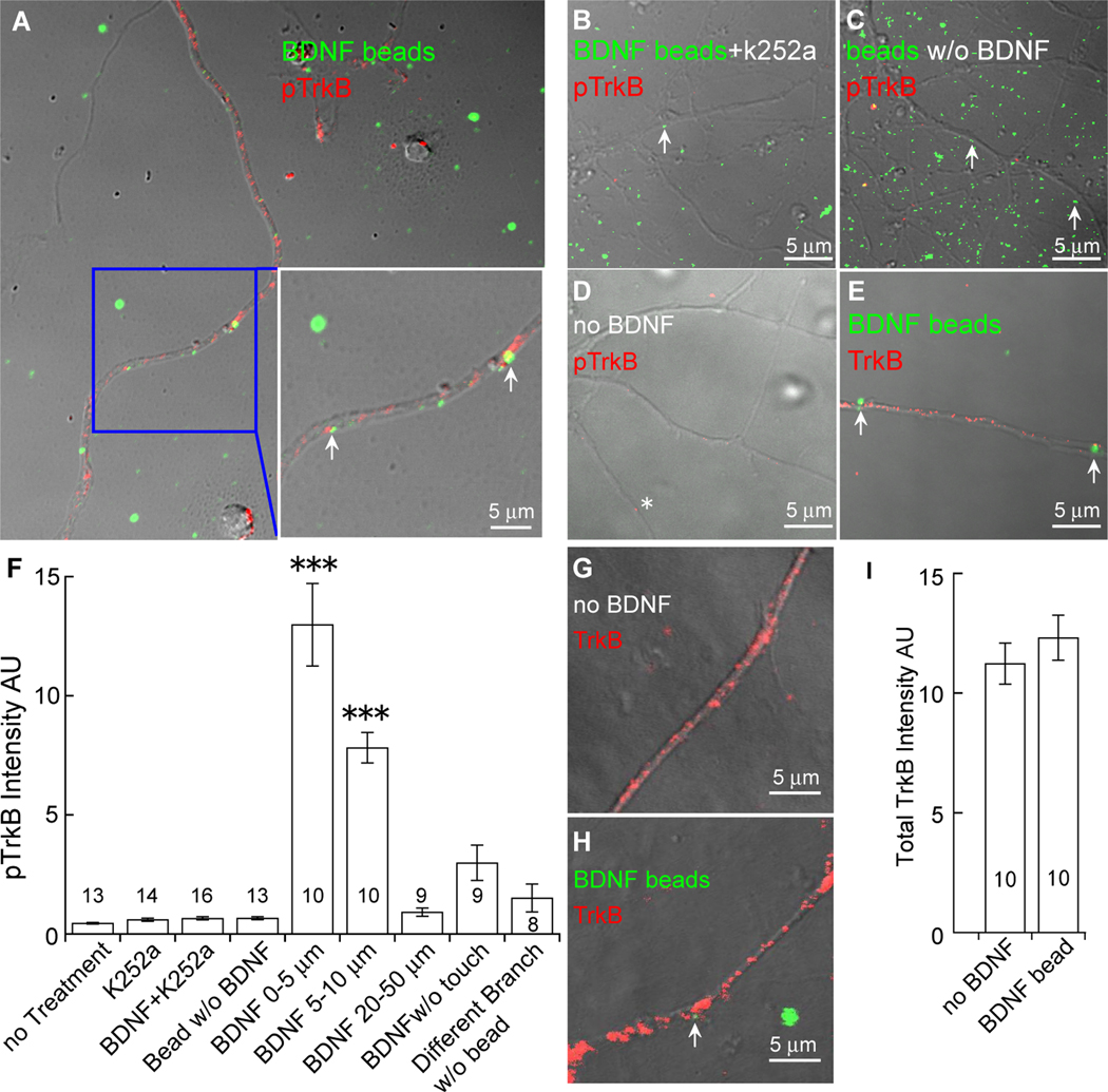
Figure 2. Local activation of TrkB by BDNF.
Hippocampal neurons were treated with BDNF-beads (green) for 30 min, and stained with an antibody against phosphorylated TrkB (pTrkB, red). Local pTrkB immunoreactivity is dramatically increased in dendrites touched by BDNF-beads (arrow).
(A) Preferential activation of TrkB on dendrites in contact with BDNF-beads. The area highlighted by the blue box is enlarged and shown in the lower right. Note the strong pTrkB signals (red) associated with the BDNF-beads (green), indicated by arrows.
(B) Blockade of BDNF-beads induced TrkB activation by k252a. Neurons treated with k252a and BDNF-beads (green) show little pTrkB immunoreactivity even when dendrites were touched by BDNF-beads (arrow).
(C) Touching by beads alone does not activate TrkB. Neurons were treated with uncoupled beads (green) without BDNF. Little pTrkB immunoreactivity was detected along the dendrites even when dendrites were touched by beads (arrow).
(D) Low levels of basal TrkB activation in cultured hippocampal neurons. Little pTrkB immunoreactivity was detected in dendrites in absence of BDNF beads (asterisk).
(E) Abundant TrkB immunoreactivity (red) along the dendrites of cultured hippocampal neurons. Arrows show the place where BDNF-bead touched dendrite.
(F) Quantification of pTrkB immunoreactivity under different conditions. The intensity of an individual pTrkB immunofluorescent spot along the dendrite was quantified by Image J. The intensities of all spots from the point touched by BDNF beads (0–5 µm, 5–10 µm, 20–50 µm) were summed and presented as arbitrary units (AU). pTrkB signal gradually decreases along the dendrites from the point of BDNF-bead contact. Note that there is little pTrkB signal on the dendritic branch not contacted by BDNF-bead, despite the fact that other branches are contacted by BDNF-beads. The measured regions of interest are indicated as “n” for each condition. ***: P<0.001; Newman-Keuls analysis following one-way ANOVA.
(G–I) Confocal images of total TrkB immunoreactivity (red) along the dendrites of cultured hippocampal neurons treated with (H) or without (G) BDNF-conjugated beads, and the quantifications (I). Note that total TrkB level with BDNF-bead treatment was comparable to control without BDNF-conjugated beads.

