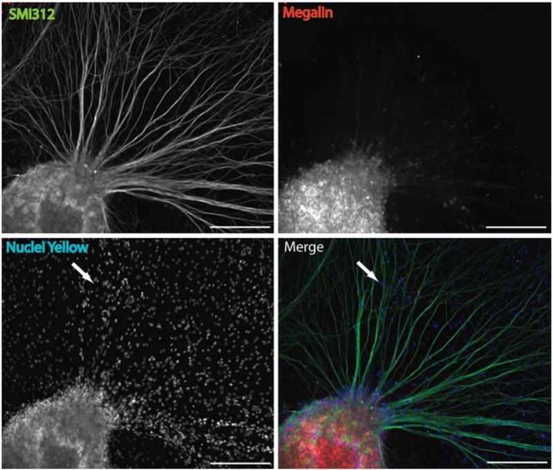
Fig. 4.

The expression of megalin in DRG explants in vitro. Immunohistochemical staining of DRG explant cultures for megalin (red) and neurofilaments (SMI312; green) revealed that megalin staining was punctate and restricted primarily to the soma of DRG neurons, with limited expression in axons. Nuclei staining (c) indicating the presence of non-neuronal cells (SMI-negative cells) within the culture (arrow in panel c and d); however, these did not express megalin. Therefore, megalin is specifically expressed by neurons in the DRG explant cultures (scale bar = 200 μm)
