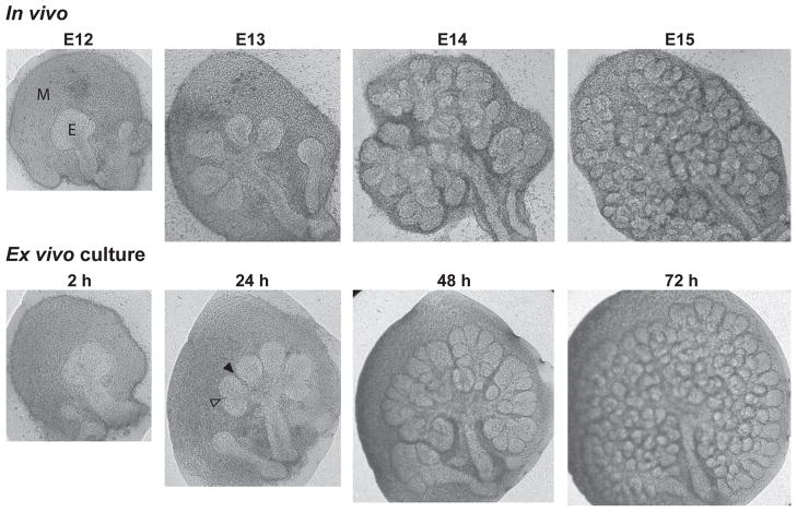
Figure 1. Branching morphogenesis of submandibular glands (SMG) in vivo and in ex vivo culture.
Top panel: Mouse SMG isolated from embryonic day 12 (E12) through E15 illustrate the formation of a highly branched structure from a single epithelial bud. Bottom panel: The pattern of in vivo branching morphogenesis can be closely reproduced by ex vivo culture of an E12 SMG. M denotes mesenchyme, E denotes epithelium, and empty and filled arrowheads show examples of early and deepening clefts, respectively.

