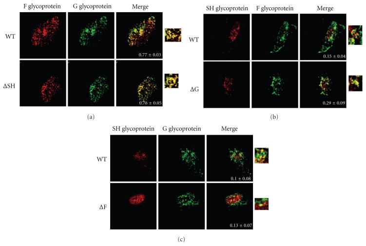
Figure 3.
Effect of the deletion of individual glycoprotein genes on the colocalization of pairs of HRSV glycoproteins at the plasma membrane. Localization of HRSV glycoproteins at the site of viral assembly by confocal microscopy. A549 cells infected with WT or glycoprotein deleted HRSV as indicated at a MOI 0.2 were fixed, permeabilized, and incubated with primary antibodies against two different glycoproteins. (a) Staining of F and G glycoproteins where F is shown in red and G in green. (b) Staining of SH and F where SH is shown in red and F in green. (c) Staining of SH and G where SH is shown in red and G in green. A merge of the two stains is shown in the right row. A blowup of the merge is also shown. Images were taken on a Zeiss confocal microscope. An average of twelve cells were imaged per staining, and colocalization was measured using ImageJ and JACoP plugin. Pearson’s coefficient ± standard error of mean is shown within the merge panels.

