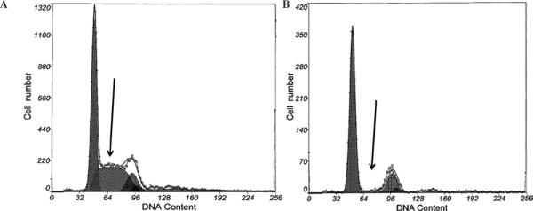
Figure 3.
The exposure of MDA-MB-453 cells to U0126 at a concentration of 10 μM at different time periods (from 15 min to 24 h) led to a dramatically reduced S-phase fraction in the cells as measured by flow cytometry. Black arrows indicate S-phase fraction after 24 h in control group (A) where S-phase was ~39%, and exposed group (B) where S-phase was ~6%.

