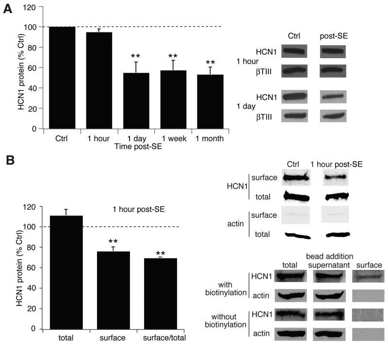
Figure 2.
HCN1 channel protein expression falls after a delay following SE, while surface expression declines immediately at 1 hr post-SE. A, HCN1 channel protein expression at 1 hr post-SE was unchanged, while at 1 d post-SE was decreased and remained reduced for at least 1 mo. Representative blots are shown in each condition, along with blots of β-tubulin III as a marker of neuronal protein content. Summary data at 1 wk and 1 mo are from (Jung et al., 2007). B, Expression of surface HCN1 channel protein was decreased at 1 hr post-SE compared to control. Control and post-SE samples were processed in the same gel to enable accurate comparison; total and surface samples were processed in separate gels and thus cannot be directly compared. Lack of actin staining in the surface fraction confirmed the absence of cytoplasmic proteins. Bottom panels from a single representative gel show that no surface protein was recovered with avidin-complexed beads when biotinylation was omitted.

