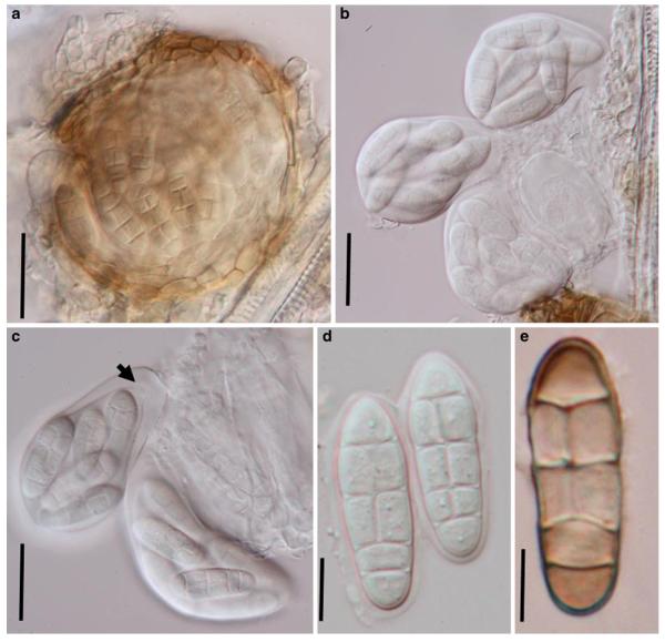
Fig. 45.
Leptosphaerulina australis (from NY, C.T. Rogerson 3836). a. Compressed ascoma. Note the obpyriform asci within the ascoma and the thin peridium. b, c. Eight-spored asci released from the ascomata. Note the apical apparatus (arrowed). d. Ascospores with thin sheath. e. An old pale brown ascospore. Scale bars: a-c=50 μm, d, e =10 μm

