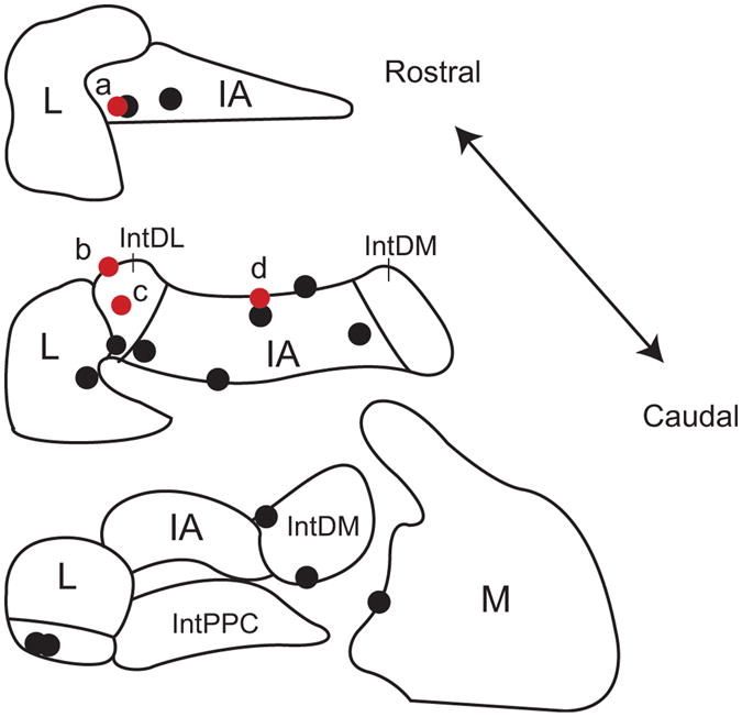
Figure 9.

Locations of recorded CN neurons. Locations of CN neurons with (red circles) and without (black circles) synaptic input from crus 2a PCs as identified by analysis of CS-CN correlograms. Schematics of coronal sections of CN based on atlas of Paxinos and Watson (1998). IA, interpositus anterior; IntDL, dorsolateral division of interpositus; IntDM, dorsomedial division of interpositus; IntPPC, posterior parvicellular division of interpositus; L, lateral nucleus; M, medial nucleus.
