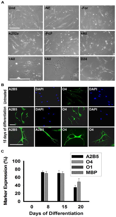
Figure 2.
Phase Contrast Images and Immunocytochemical Analysis of Differentiating MLPCs in 2D Environment. (a) Undifferentiated MLPCs (Und) exhibited fibroblast morphology. Cells at 15 days in differentiation medium without NE, forskolin (For) or K252a, retained their fibroblast morphology. Process formation was visible in the medium without growth factors PDGF-AA, EGF and bFGF (PEF). Refractile cell bodies and increased process formation were observed in the presence of all factors indicated above for days 15–20. Cells lost their multipolar morphology and became bipolar or spindle shaped after about day 20–24. Scale bars, 100 μm, (20x and 40x magnification). (b) Immunocytochemical analysis of differentiating MLPC in 2D environment. The untreated MLPCs showed negative staining for A2B5 and faint staining for O4. At 15 days of differentiation, cells exhibited positive staining for A2B5 and O4, characteristics of immature oligodendrocyte precursor cells. Scale bars, 100 μm, (Rows 1 and 2, 20x magnification and Row 3, 40x magnification). (c) MLPCs do not differentiate into committed oligodendrocytes in 2D environment. At 8 days of differentiation, 72.4% of cells were positive for A2B5 and 69.9% for O4 and at day 15 70.3% of MLPC exhibited positive staining for A2B5 and 69.7% for O4. At 20 days, 35.0% of cells remained A2B5 positive and 49.7% O4 positive. Expression of O1 galactocerebroside and MBP was absent in both untreated and differentiating cells. Error bars represent the SD.

